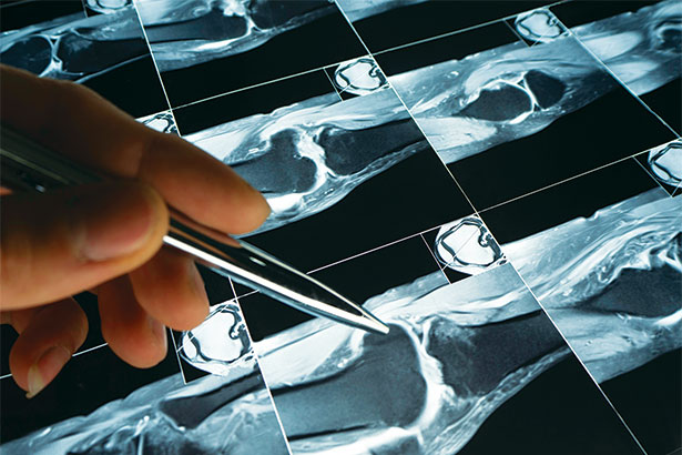
The latest imaging technology is changing clinical care at Englewood Health, leading to faster, more accurate diagnoses of musculoskeletal disorders.
While traditional MRI scanners use magnetic fields measuring 1.5 tesla, the 3-tesla MRIs at Englewood Health use magnets with twice the power to create more highly detailed images. This enhanced resolution enables referring physicians to make diagnoses that may have been missed with conventional imaging.
As Mark Shapiro, MD, the chief of radiology at Englewood Health explained, with conventional MRI scanners, a small portion of patient anatomy is difficult to see due to limited sensitivity and resolution of the scanner. In contrast, the scanners at Englewood Health offer up greater resolution, giving referring physicians and orthopedic surgeons far more confidence in the diagnosis.
“The greatest advantage is in the smaller joints like the wrist, hand, foot or ankle,” Dr. Shapiro said. “The higher strength magnets enable us to see these tiny structures with enough detail that we can diagnose most abnormalities when they appear.”
The machines are fast, too—up to 25% faster than the previous generation of MRI scanners. A quicker examination is not only easier for patients, who might be asked to hold an awkward position for 30 minutes or longer, but the speed also increases accuracy.
“Motion is your enemy in the MRI,” Dr. Shapiro said. “By utilizing increased speed, we’re eliminating motion artifacts that are very common.”
“Motion is your enemy in the MRI,” Dr. Shapiro said. “By utilizing increased speed, we’re eliminating motion artifacts that are very common.”
Mark Shapiro, MD
Although 90% of musculoskeletal imaging is geared toward use of MRI, Dr. Shapiro and his radiology team, composed of subspecialty fellowship-trained radiologists, also employ ultrasound imaging for nonsurgical procedures, such as anesthetic and corticosteroid joint injections, applied directly to damaged joint tissue.
“Ultrasound imaging is easier for patients, and we’re able to perform therapeutic procedures without exposing them to radiation,” said Dr. Shapiro, who noted that fluoroscopy was previously used to guide the needle into the joint.
Ultrasound is also used for the diagnosis of tendon or ligament tears and the characterization of mass lesions in patients who cannot undergo MRI (e.g., because of a pacemaker, claustrophobia or body habitus).
April 14, 2021

