
MRI and ultrasound combine to provide enhanced view of prostate
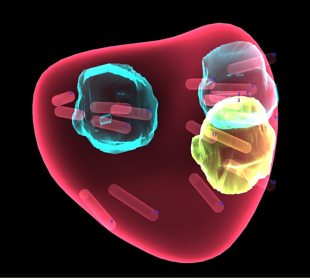
An advanced technology called MRI ultrasound fusion biopsy is now being used to diagnose prostate cancer at Englewood Hospital and Medical Center. The technology uses a two-step process that offers greater accuracy over traditional prostate cancer biopsies. First, a patient undergoes magnetic resonance imaging (MRI) of the prostate. A radiologist evaluates the images and identifies and marks any suspicious areas for further evaluation by a urologist. The urologist then fuses or superimposes the images with real-time ultrasound images, which produces 3D images of the suspicious areas and offers improved ability to biopsy the prostate.
Some 60 percent of newly diagnosed prostate cancer patients are considered low-risk on initial biopsy, but if a biopsy is repeated, that number drops to 30 percent. Those considered to be at low risk can be placed on active surveillance, or watchful waiting, but standard biopsy has shortcomings in determining the risk level. This new MRI ultrasound fusion biopsy technology has been shown to more accurately determine whether a patient has higher risk prostate cancer or lower risk prostate cancer. By risk stratifying patients, physicians can better inform them about their treatment options, be it active surveillance or surgery or radiation therapy.
“This is an exciting advancement in imaging and cancer diagnosis,” said Dr. Mark Shapiro, chief of radiology at Englewood Hospital and Medical Center. “Using a 3-Telsa magnet – the most advanced on the consumer market – MRI is proving to be effective in diagnosing clinically significant prostate cancers that require treatment. As a radiologist reading the images, I can now localize for the urologist a potentially clinically significant lesion that requires biopsy. MRI by itself cannot diagnose prostate cancer, but it is a powerful tool to help urologists provide the most accurate diagnosis for patients to give them a personalized treatment plan.”
The traditional approach to prostate biopsy involves ultrasound, but without the benefit of enhanced visualization offered by the fusion technology. Using an ultrasound probe with a needle through the anus, a urologist extracts small samples of prostate tissue to look for cancer. But these ultrasound images are generally of poor quality. As a result, the samples are taken from random areas of the prostate and can miss a prostate tumor entirely.
MRI ultrasound fusion biopsy is ideal for men who have an elevated prostate-specific antigen (PSA) as determined by a blood test, including those with a previous negative biopsy result, men with an abnormal rectal exam, or men diagnosed with prostate cancer who are on active surveillance, also known as watchful waiting.
The procedure is performed on an outpatient basis. A patient must first be evaluated by a urologist to determine if he is a candidate for MRI ultrasound fusion biopsy. For more information and a referral, patients can contact The Lefcourt Family Cancer Treatment and Wellness Center at Englewood Hospital and Medical Center 201-608-2266 or cancer.center@ehmchealth.org.