
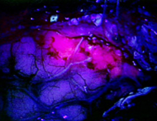
Fluorescing tissue “lit up” by Gleolan™ facilitates maximal and safe removal of glioma cells from brain tissue during surgery.
April 25, 2019 — Englewood Health is the first hospital in New Jersey to use a novel fluorescent technology that makes brain cancer cells “light up” when viewed through a special microscope. The innovative optical imaging agent—called Gleolan™—enables surgeons to see and remove malignant glioma cells remaining in brain tissue during surgery, while also avoiding parts of the brain responsible for vital functions such as speech and movement.

“This is game-changing technology for glioma surgery,” explained Kevin Yao, MD, the Englewood Health neurosurgeon who performed the first procedure in early March. He removed a four-centimeter malignant glioma from a woman in her 70s who was experiencing headaches and confusion and had noticed changes in her speech prior to receiving her diagnosis.
High-grade gliomas are highly challenging brain cancers to treat successfully and are not always operable. The tumor often comes back after treatment. So techniques that permit the surgeon to remove as much tumor tissue as possible could enhance a patient’s outcome.
Gleolan is a medication called aminolevulinic acid HCl. The patient drinks the medication two to four hours before receiving anesthesia for the surgery. During the operation, the neurosurgeon views the brain through special blue light filters on a surgical microscope. Under this blue light, Gleolan helps tumor cells to “fluoresce,” glowing a red-violet color. The medication is designed to make cancerous brain cells glow, but not noncancerous cells. Gleolan was approved by the U.S. Food and Drug Administration in June 2017 for use during surgery for high-grade gliomas, and is commercially available to all patients.
During the procedure at Englewood Health, Dr. Yao removed the tumor tissue he could see with the naked eye. He then viewed the area through the microscope and could see that there were cancer cells remaining, and was able to remove those as well. “It was empowering because I could actually see the tumor cells and was able to remove more during surgery than I would have been able to without this technology,” he noted.
The technique adds to the arsenal of tools that surgeons use to “map” the location of a brain tumor. Current techniques provide a rough idea of a tumor’s location. However, brain anatomy can change during an operation as the tumor is removed and tissue shifts. Intraoperative MRI is also used to map brain tumor location, but it is costly and resource-intensive, requiring all equipment in the operating room to be MRI-compatible. Unlike existing brain mapping techniques, Gleolan provides real-time information showing exactly where glioma cells are lurking in the brain.
Dr. Yao added that Gleolan, combined with functional imaging and testing, showed him that the patient’s tumor tissue was near, but not in, areas of her brain responsible for speech. “Every extra millimeter of tissue that I remove can improve her survival, but can also dramatically affect her speech. Gleolan allowed me to maximally and safely remove the tumor,” he explained. “I think this approach is going to change the standard of care for glioma surgery.”
Fluorescing tissue “lit up” by Gleolan™ facilitates maximal and safe removal of glioma cells from brain tissue during surgery.