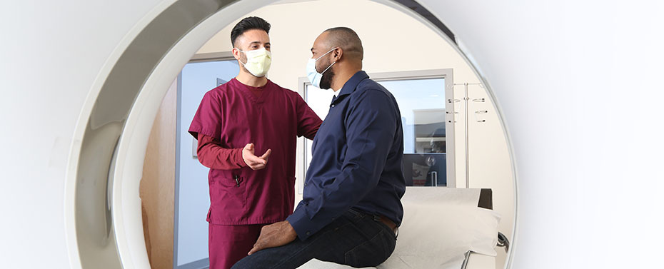
Our Department of Radiology is committed to serving the community with the best equipment, an unsurpassed staff, and convenient, exceptional service. We offer testing onsite at Englewood Hospital and Medical Center and at two offsite locations: Advanced Medical Imaging (AMI) in Emerson, New Jersey (map), and Magnus Imaging in Glen Ridge, New Jersey (map). We hold accreditation in MRI, CT, ultrasound, nuclear cardiology, and mammography from the American College of Radiology. Englewood Hospital and Medical Center is also home to a School of Radiography to develop entry-level radiographers to serve the community.
We offer extended hours and same-day appointment availability. To make appointment, call 201-894-3641 or book online now.
Care & Treatment
Locations and Services
Englewood Hospital and Medical Center
350 Engle Street
Englewood, NJ 07631
201-894-3400
To make appointment, call 201-894-3641 or book online now.
Free self-parking and valet parking.
Services:
- X-Ray
- MRI
- Mammography
- Low-dose CT scan
- PET scan
- Nuclear cardiology
- Interventional radiology
- Ultrasound
Hours:
- Weekdays 8:00 a.m. – 8:00 p.m.
- Weekends and holidays 8:00 a.m. – 4:00 p.m.
Advanced Medical Imaging (AMI)
452 Old Hook Road
Emerson, NJ 07630
201-262-0001
To make appointment, call 201-262-0001 or book online now.
Hours:
Monday – Thursday: 7:30 a.m. – 8 p.m.
Friday: 7:30 a.m. – 5:30 p.m.
Saturday: 8 a.m.- 2 p.m.
Services:
- X-Ray
- MRI
- CT/CAT scan
- Ultrasound
- Mammography and other breast imaging and diagnostic services, including breast ultrasound, clinical breast exam, stereotactic breast biopsy, and tomosynthesis.
Magnus Imaging
946 Bloomfield Avenue
Glen Ridge, NJ 07028
To make an appointment, call 973-743-0061
Services:
- CT/CAT scan
- Nuclear cardiology
- Echocardiology
- Vascular ultrasound
Leslie Simon Breast Care and Cytodiagnosis Center
350 Engle Street
Englewood, NJ 07631
201-894-3202
To make appointment, call 201-894-3202 or book online now.
Services: Visit the Breast Center website to learn more about the services.
Diagnostic X-Ray
Diagnostic radiology procedures use radiation to help identify disease or injury. X-rays penetrate body tissues and produce an image that provides vital information about normal structures as well as diseases in the body. Diagnostic X-rays are used to examine all parts of the body. For example, diagnostic X-rays can be used to identify problems in the digestive system and kidneys, and find evidence of bone fractures and dislocations.
Typically, patients will be asked to lie down on an X-ray machine and hold still while the X-rays are taken. For most patients, diagnostic radiology is a quick and painless procedure. Depending on the tissues or organs being studied, the procedure may involve the use of a contrast medium, which will help make soft tissues stand out. For example, in an arthrogram, a test used to examine joints, a contrast medium is injected into a joint to detect injury or disease. If you need an upper or lower Gl series, used to examine the digestive tract, you may be asked to drink a contrast material or it may be administered by enema.
Your doctor will provide detailed instructions on preparing for the test. Preparations will vary depending on the procedure. For example, patients may be asked to make dietary changes or avoid food or drink for a period of time prior to having the examination. Patients who are pregnant must alert their doctor, who will decide whether it is safe to have an X-ray examination. Doctors should also be advised of any allergies or any previous reactions to a contrast medium. You should wear comfortable clothing and leave jewelry at home.
Magnetic Resonance Imaging (MRI)
Magnetic resonance imaging, or MRI, uses radio waves and a powerful magnetic field to produce cross-sectional images of internal body structures. An MRI is a painless procedure that can clearly map out soft tissue, producing images that are extremely precise without exposing patients to radiation or radioactive solutions. An MRI can provide physicians with vital information to diagnose diseases or disorders so that proper treatment can be initiated promptly, often eliminating the need for more invasive procedures. MRI is particularly valuable in studying the brain and spinal cord diseases; revealing and diagnosing tumors and metastatic diseases; providing detailed images of the cardiovascular system; examining joints and soft tissues; imaging abdominal organs – especially any abnormal masses or aneurysms; detecting abnormalities of the neck, abdomen and pelvis; and identifying musculoskeletal problems.
Englewood Hospital and Medical Center is home to a 3 Tesla MRI machine, one of the most advanced tools of its kind. When compared to the traditional 1.5T MRI system, the 3T delivers twice the magnetic field strength, which results in higher-resolution images. The machine is especially helpful in neurosurgery and orthopedics. A wider, open design helps reduce feelings of claustrophobia and accommodates patients weighing up to 550 pounds.
MRI is a painless procedure. Most scans take about 45 minutes. Our technologists
will explain the procedure and remain to answer any questions. Patients must lie still for the duration of the test on a comfortable padded table. The short-bore magnets allow patients the comfort of gently gliding into the MRI’s short, flared opening cylinder, thus usually eliminating the claustrophobic effects of longer magnets. Sometimes an injection of a contrast material is required. Patients are encouraged to discuss concerns about the test with their doctor and the technologist.
There is usually no special preparation necessary before having an MRI. Food and drink intake is typically not restricted before the procedure. However, patients will be asked to remove all metallic objects, such as jewelry, glasses and clothing with zippers, before the exam.
Some people should not receive an MRI. People with metal implants, such pacemakers, aneurysm clips and implanted stimulating devices, cannot have an MRI. Patients should discuss these health issues with their doctor.
Patients should wear comfortable clothing without zippers, such as a sweatsuit, wear little or no makeup, and leave jewelry at home.
CT/CAT Scan
A CT (or computerized tomography) scan combines computerized technology with a focused X-ray beam that passes through the body at various angles, producing clear, cross-sectional images. It is safe, fast and painless, and produces precise and accurate images of the body. A CT scan is one of a physician’s most important tools for examining the bones and internal organs. CT scans are able to detect many conditions that conventional X-rays cannot. CT scans can often take the place of other more invasive diagnostic techniques, such as exploratory surgery, saving the patient discomfort, cost, and inconvenience. The scans are also useful for monitoring a patient’s progress during or after treatment. CT scanning is considered a critical component of any evaluation of brain injuries or function. CT scans of the head may also be used to detect or rule out tumors, blood clots, and other disorders. These scans can also be especially important in diagnosing enlarged lymph nodes, pancreatic disease, back problems, or lung cancer. CT (or computerized tomography) is also used to perform a virtual colonoscopy, a procedure that examines the colon using conventional CT techniques.
Low-dose lung cancer CT screening is a relatively new procedure performed with CT (or computerized tomography). A CT scan combines computerized technology with a focused X-ray beam that passes through the body at various angles, producing clear, cross-sectional images of the lungs to check for lung nodules that may be cancerous. This procedure is safe, fast, and painless. This screening is recommended for patients with a history of smoking. Studies have shown that a CT scan is much more sensitive than a conventional chest X-ray in detecting lung modules that may be cancerous.
The Centers for Medicare & Medicaid Services (CMS) has determined that the evidence is sufficient to cover this screening if you have Medicare, are 55-74, a current or former smoker, and meet additional criteria. A prescription from a physician is required.
The procedure is fast and painless. Patients will be asked to lie on a table that will move through the scanner opening, taking images of the body. Patients may be asked to remain very still and hold their breath while the actual images are being made. The whole procedure usually lasts 30 minutes, depending on the type of test being performed.
Doctors will provide detailed information on preparing for the scan. Patients may be told to avoid food or drink for around 4 hours before the test, especially if an intravenous contrast injection is to be used. An oral contrast medium is often given, as well as a contrast enema.
These techniques are used to highlight certain structures, including the stomach and bowel, making the images more diagnostic. Some people are allergic to certain contrast media.
Patients with allergies, particularly to iodine, should inform their doctor and the medical center staff ahead of time. Patients who are pregnant must alert their doctor, who will decide whether it is safe to have a CT scan.
Patients should wear comfortable clothing, leave jewelry at home, and must remove hairpins, eyeglasses, and dentures when having a head scan.
PET CT Scan
This examination combines two scanning techniques into one — the PET (positron emission tomography), which shows different functions in the body, and the CT (computed tomography), which shows the detailed structural anatomy. This combination allows doctors to get highly defined 3D images from the inside of the body with only one system and during one appointment. It combines both functional and anatomical information into one single scan. Your doctor will be able to pinpoint the exact location of interest and determine its functional status. One of the most important factors in the fight against any disease is early detection. A PET/CT scan provides your doctor with information that may help to specify and improve any treatment that you require and possibly even reduce the risk of surgical procedures.
A PET/CT examination is straightforward and painless. You will receive a small injection of a harmless radiolabeled sugar called FDG. This will circulate through your body and will be metabolized or “used” by the cells and organs.
Some cells and many diseases use this sugar more than others, and the PET system can measure these differences. For this reason, after the small injection, you will be asked to relax for about an hour while the sugar circulates. Afterward, you will be asked to lie down on the scanner bed. Simply relax and follow the technologist’s instructions. It is very important to remain as still as possible during the exam and breathe normally unless told otherwise. You may be required to keep your arms over your head during the exam.
The scanner bed will move in toward the scanner opening for a short time, during which you will hear some noise coming from the system. This will complete the CT portion of the scan. Next, the PET scan will begin. The bed will once again move, but not as quickly. The technologist will keep you informed about what is going on. Although tissues in the body will normally process FDG, diseased cells process the FDG more rapidly than healthy tissues. It is this increased absorption of FDG that is captured by the PET scan and determines if there are any problem areas. When the PET and CT images are combined, it completes the entire picture and helps your physician in making an accurate diagnosis.
Patients will be provided with detailed instructions on scans from their physicians. Some people, such as women who are pregnant or breastfeeding, should not receive a nuclear medicine scan. Patients should discuss any health issues that may rule out the use of nuclear scans with their physicians. Depending on the test being done, patients may be asked to avoid food and/or water for a period of time before the test. A change in diet or medications taken in the weeks before the test may be required.
Ultrasound
Ultrasound scanning is a diagnostic technique in which very high-frequency sound waves are used to “see” inside the body. Also called sonography, ultrasound emits sound waves that can be bounced off (echo) the kidneys, liver, heart, or other soft tissue and converted into images on a television screen. No radiation is involved in this procedure. Ultrasound is a versatile and comfortable method of diagnostic testing that can provide excellent images of the body and its organs. It can also be used to analyze blood flow through vessels throughout the body. During pregnancy, ultrasound can evaluate the growth, health, and position of the developing fetus.
Englewood Hospital and Medical Center offers pregnant patients the option of having ultrasound exams performed in the Antepartum Testing Center. This department is solely dedicated to the concerns of pregnant women and fetal evaluation.
In general, patients can expect to change into a hospital gown. The ultrasound technologist or physician will apply a water-soluble jelly to the area of the body being examined and the ultrasound transducer will be passed over the area. Images will be formed by a computer and displayed on a video screen.
Sometimes, special preparation is necessary. Depending on the type of ultrasound scheduled, patients may be asked to drink several glasses of water prior to the test or refrain from eating approximately 8 hours prior to the examination.
Uterine Fibroid Embolization
Uterine fibroid embolization (UFE) is a minimally invasive procedure used to shrink fibroids, which are benign (non-cancerous) tumors that develop in the muscular wall of the uterus. UFE can reduce or eliminate symptoms of fibroids, including heavy menstrual bleeding, pelvic pain and pressure, back or leg pain, painful sexual intercourse, bladder pressure leading to a constant urge to urinate, or pelvic fullness or abdominal bloating. UFE relieves these symptoms in 90 percent of patients.
Englewood Hospital and Medical Center’s vascular interventional radiologists are specially trained to perform UFE. The procedure is also used to treat obstetrical hemorrhage and other bleeding problems in the pelvis. Nationally and internationally recognized for our expertise in transfusion-free medicine and surgery, we ensure patients minimal use of blood products in all treatments for fibroids. In addition to UFE, we have other nonsurgical and surgical treatment options for the shrinkage and removal of uterine fibroids.
UFE requires an overnight hospital stay. Most procedures take 60 to 90 minutes, and you will be sedated and remain drowsy. An interventional radiologist inserts a small catheter into the femoral artery in the groin or radial artery in the wrist. An X-ray is performed to give the doctor a clear picture of the blood supply to the uterus and fibroids. The catheter is guided through the artery to the uterus and tiny particles are slowly injected into the arteries that supply blood to the fibroid. These particles block the blood supply to the fibroids, shrinking them by up to 65 percent. Although blood flow to the fibroids is blocked, the artery will continue supplying blood to the normal portions of the uterus. A local anesthetic is used to numb the skin and no stitches are needed after the procedure.
After the procedure, many patients experience several hours of moderate to severe pain, fever, and nausea, which can be controlled with intravenous medications. By the next day, most symptoms improve and patients can go home. Many patients experience several days of moderate to severe cramping, tiredness, fever, or periodic nausea. Pain medications are prescribed to control these symptoms. Most women can return to work 7 to 14 days after the procedure.
Adverse reactions occur in less than 3 percent of patients. Though uncommon, serious potential complications include injury to the uterus from decreased blood supply or infection. Hysterectomy to treat such complications occurs in less than 1 percent of patients.
The expected average reduction in the volume of the fibroids is 50 percent in three months, with reduction in the overall uterine volume of about 35 percent. Thousands of patients have had this procedure worldwide, with results showing that symptoms improve in 90 percent of patients.
Most patients who have UFE are past their childbearing age or not interested in becoming pregnant. As a result, it is unclear what effect blocking the uterine arteries will have on the ability to become pregnant or to carry a pregnancy to term. However, more than a dozen successful pregnancies have been reported by patients who have had uterine arteries embolized for other reasons, such as bleeding after childbirth. Research is underway to study this issue.
Interventional Radiology
Interventional radiologists use minimally invasive image-guided procedures to diagnose and treat diseases in nearly every organ system. Among our interventional radiology services are angiography, chemoembolization, y90 procedures, uterine ablation, microwave ablations, and more.
Angiography
Angiography is a procedure that enables blood vessels to be visualized after they have been filled with a contrast medium. Angiography can be coupled with certain types of procedures called interventional radiology. Interventional radiology techniques can repair blockages in the vessels, such as cholesterol build up, without performing surgery. The most common type of interventional radiology is the balloon angioplasty, in which a small balloon is inserted into a blood vessel and inflated to expand a narrowed or blocked segment of an artery or vein. Angiography and angioplasty are often recommended for patients diagnosed with peripheral vascular disease, a condition in which plaque (fatty cholesterol) builds up in the blood vessels and impedes the flow of blood. Angioplasty is less invasive than surgery and performed under a local anesthetic. The entire procedure takes about one hour.
What to expect during angiography
The first step in angiography is usually the diagnostic X-ray of the blood vessels to identify the narrowed blood vessel. This procedure can be followed by an angioplasty to treat the blockage in the vessel.
During the procedure, the patient lies flat covered with a sterile drape. An area of the arm or leg will be disinfected with liquid soap. After a local anesthetic has been applied to numb the area, a small plastic tube, or catheter, is inserted in into an artery of the arm or leg. Following insertion, the catheter can then be directed through the blood vessels to the part of the body under evaluation. Once in position, X-ray contrast (dye) is then injected through the blood vessels and special X-rays called arteriograms are taken to identify the diseased area.
An angioplasty may be performed through the same tube used for the arteriogram; sometimes a second tube is inserted for the balloon. A special instrument with a deflated balloon is inserted through the tube and positioned at the narrowed area. The balloon is inflated to open the narrowed or blocked section of the vessel. Sometimes, a special metal stent is inserted to reinforce the walls of the vessel. This stent is incorporated into the vessel where it will remain permanently.
Following this procedure, the patient may be required to stay in the hospital overnight for observation. Most patients are able to walk the next day and return to their normal routine within two to three days. In fact, most patients’ symptoms are significantly improved by the procedure and they are able to resume greater activity than prior to the procedure.